


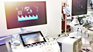
Ultrasound scans are one of the most common medical procedures today. With the help of ultrasound exams, deeper imaging is possible than with X-rays, without any harmful health concerns.
Even though most people have heard of ultrasound medical imaging, not many know all its applications. Ultrasound machines are in almost every medical department.
In this article, we will discuss the applications of ultrasound machines in medical science.
Overview of Ultrasound Technology
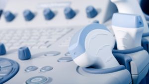
Ultrasound technology uses high-frequency sound waves that human hearing can’t detect. A typical ultrasound beam is transmitted by a control unit at a speed greater than 20,000 Hz.
There is a frequency shift as the wave travels through different densities of materials in the body caused by their different mechanical properties. And these changes are recorded by digital processors and transformed into an ultrasound image.
The body part to be scanned/examined determines the frequency of sound waves selected for imaging purposes. The process uses electrical pulses emitted by a transmitter pulse generator. The piezoelectric transducer converts these electrical pulses into vibrations that create sound waves.
Application and Uses of Ultrasound in Medicine
Here is a detailed breakdown of the use cases of ultrasound scanning in medicine:
Abdominal
- An ultrasound device is ideal for diagnosing the cause of any abdominal pain or discomfort. It can scan and identify problems with soft tissues in the abdomen that are not functioning correctly.
Nephrology
- Ultrasound scanning is the only technology applied to detect the presence of kidney stones. It can even detect the number and size of each stone in real time.
- A renal lithotriptor using ultrasonic waves can also break kidney stones. This process is known as lithotripsy. With lithotripsy, pulses of ultrasound break large kidney stones into smaller fragments. The smaller fragments can easily pass through the urinary tract.
Musculoskeletal
- Ultrasound examinations allow doctors to see the blood vessels, muscles, and joints inside the body. The images help investigate muscle pulls, tears, nerve problems, and ailments such as arthritis and osteoporosis.
- Ultrasound is used to treat soft tissue ailments, bursitis, collagen diseases, and soft tissue injuries. Decreasing pain and reducing soft-tissue stiffness results in a shorter healing time.
Cancer
- With the help of ultrasound elastography, medical professionals can detect tumors in patients that are not visible with other medical imaging technologies. Ultrasound-guided biopsies are normal.
- One of the extensive applications of ultrasonic waves is breast examinations and the study of breast cancer. Medical ultrasound imaging help detect the lumps in the breast and guide the needle in taking the sample of this lump for further analysis.
- Apart from ultrasound elastography, the ongoing development of using ultrasonic hyperthermia to treat cancer is also promising. Ultrasonic hyperthermia increases the temperature of the target area above a particular level. At this high temperature, halting the malignancy of cancer is expected.
Surgery
- A high-energy surgical ultrasonic device can destroy tissue intentionally. In some surgical cases, a specialized setup has multiple piezoelectric transducers focused on the site tissue while avoiding any intervening tissue.
- Another use is in the surgical treatment of Parkinson’s disease by the ablation of substantia nigra.
Urology
- Ultrasound exams are used to examine the urinary tract for any defects. It can also detect urinary tract infections and problems with voiding.
- Many dedicated types of equipment have been developed utilizing ultrasound technology. These are very successful in the treatment of Meniere’s disease, which causes improper inner ear functioning and makes the patient feel vertigo.
- Conventional surgery has a high-risk factor of deafness when treating Meniere’s disease. Ultrasound eliminates this risk, stops vertigo, and decreases the risk of facial nerve paralysis.
Gynecology
- An ultrasonography examination is used to study the female pelvic organs, mainly the ovaries, fallopian tubes, uterus, bladder, adnexa, and recto-uterine pouch.
- It can detect inflammation of the appendix (appendicitis). Ultrasonography can also detect endometriosis, ovarian cysts, lesions, and gynecological cancer.
Ophthalmology
- A phacoemulsifier is used for surgically treating cataracts in the eyes. This surgical instrument uses a needle vibrating at an ultrasonic frequency. The high energy released by the vibrations acts as an emulsifier for the dead lens. A phacoemulsifier also contains a suction chamber that removes the debris created in the process, providing a faster surgery time and recovery period.
Anesthesiology
- Critical Care Ultrasound is used while administering anesthesia to study the blood flow. Doctors use Transesophageal Echocardiography (TEE) to determine the depth of epidural space in patients with difficult anatomy.
Endocrine

- Using an ultrasonic beam to create 3D images can help identify abnormalities, such as thyroiditis, in thyroid and parathyroid glands. The 3D images show the dimensions of the glands to identify any swelling.
Gastroenterology
- In gastroenterology, an ultrasonic endoscope helps diagnose problems with the digestive tract and soft tissues and organs in proximity. Evaluating the digestive organs and diseases in them does not require incisions with this method, making it a minimally invasive procedure.
Obstetrics and Pregnancy
- Obstetrics is one of the most common applications. It is one of the primary reasons that many people are familiar with ultrasound imaging examinations.
- During pregnancy, fetal ultrasounds provide a high-quality two-dimensional image of the embryo or the fetus in real-time. Since X-rays are dangerous for the fetus, fetal ultrasounds offer a safer method to monitor a pregnancy.
- Within 18 to 22 weeks of gestation, fetal ultrasounds can detect any abnormalities in the growth or any other problems associated with the pregnancy.
- Many different types of obstetrics sonograms exist. Some common ones are 3D sonography which creates a 3D visual of the fetus, and Doppler ultrasounds which enable hearing a child’s heartbeat.
Vascular
- Ultrasounds used for vascular patient examination help evaluate the direction of blood flow and the condition of the blood vessels. A cerebrovascular examination can measure blood flow velocity in the brain using a small sample volume.
- Carotid ultrasounds detect any arteries that are blocked or narrowed. This data helps in detecting the possibility of a stroke before any mishap.
Pulmonology
- The study of thoracic diseases uses pulmonary ultrasounds. In emergency departments, pulmonary ultrasounds also monitor respiratory diseases.
- Imaging lungs can be somewhat challenging because ultrasounds work on the passage of sound waves. In the case of the lungs, air pockets can alter the behavior of sound waves and hinder the formation of optimal imaging.
Cardiology

- Echocardiography uses standard or Doppler ultrasound to produce an image. The image is usually called an echocardiogram, echo, or cardiac echo.
- With a cardiac ultrasound, it is possible to diagnose any heart disease in a patient. Echocardiograms help monitor those already suffering from heart diseases.
- The information provided by a cardiac ultrasound is extensive, so much so that this is usually the sole imaging test required for an in-depth study of the patient’s heart. It provides details about the size of the heart, its shape, any possible tissue damage with its location and size of the damage, pumping capacity, jet of blood flow, and a lot more.
- With a cardiac ultrasound, doctors can diagnose myocardial infarction early and see the regional wall motion abnormality. For patients with heart failure, it is one of the most essential tools used in the treatment.
- An echocardiogram can diagnose cardiomyopathy, dilated cardiomyopathy, and hypertrophic cardiomyopathy. Doctors also use echocardiography to identify if heart disease is the cause of chest pain in a patient.
- Technicians require training in echocardiography. Such technicians are called echo sonographers or cardiac physiologists.
Mammography
- X-rays used to screen for breast cancer in examinations of women is a process called mammography. However, the high radiation of X-rays causes a health concern for women, especially during pregnancy.
- Breast ultrasounds provide a better and safer way to examine breasts for unusual masses that could be cancerous. These are becoming the preferred method for such examinations.
- Biopsy of the breast is almost always ultrasonic-guided, guiding the probe to remove the portion of the soft tissue to examine.
Veterinary
- Ultrasounds are not just limited to the science of human anatomy. Veterinary medicine also utilizes ultrasounds in a similar way to human medicine.
- Since ultrasounds are a non-invasive imaging technique, they help save animals from undue stress caused by conventional surgeries. For many animals, surgical distress can cause additional ailments.
- It is common for household pets to ingest foreign objects. With the help of ultrasounds, the vet can detect the type of object a pet has swallowed and any danger it poses. The vet can then decide on the best course of action.
- Ultrasounds can monitor high levels of liver enzymes, which can be a common problem in many animals. Other detectable ailments are urinary tract infections, digestive disorders, neoplasia, trauma, endocrine disorders, and investigating fever of unknown origin.
- Ultrasounds can distinguish fluid, pieces of healthy tissue, and foreign objects. This distinction is impossible with alternatives such as X-rays.
- For animal gestation, vets can determine the number of offspring with ultrasonic scans. Monitoring fetal development during the entire gestation period is also easy.
Other applications
- Compression sonography can help to treat conditions such as deep vein thrombosis and injury to veins.
- Quantitative sonography can help to estimate measures of muscle quality.
Advanced Applications
- Ultrasonic imaging offers many benefits without any risks or incisions. So, experts have developed many advanced ultrasonic applications using sophisticated devices, including portable ultrasound machines, contrast-enhanced ultrasound, and more.
- One of these developments is High-Intensity Focused Ultrasound (HIFU). HIFU provides biological effects such as local tissue heating, radiation forces, and cavitation. HIFU opens new possibilities in applications such as tissue ablation, local drug delivery, immune response stimulation, and radiation therapy sensitization.
- High spatial precision is essential for these advanced treatments. This precision comes from using specialized ultrasonic transducers. These transducers can focus the ultrasound waves on an area as small as 1 mm, in diameter.
- HIFU can also treat uterine fibroids and essential tremors. Pain relief benefits of this technology are also finding applications along with research for new possibilities.
- The use of HIFU for treating prostate cancer is also under development. This method uses a HIFU transducer placed in the rectum or the urethra.
- Ultrasound molecular imaging has emerged as a powerful tool for visualizing and characterizing disease processes at the cellular and subcellular levels. This technology utilizes microscopic bubbles injected into the bloodstream targeting specific cells or tissue types.
- For drug delivery, a technique called sonoporation is gaining popularity. This technology uses microbubble-enhanced ultrasonic waves. The microbubble core contains the drug. Sometimes, an indirect drug delivery method co-administers the microbubble and drug. In all of these cases, the ultrasonic probe helps by directly guiding the drug to the pathological site.
What Are the Limitations of Ultrasound in Medicine?
While there are countless benefits of implementing ultrasounds in all the medical applications mentioned above and no adverse biological effects, there are also some limitations to the process.
- The performance of ultrasonic waves is unsatisfactory in air. Therefore, they are unsuitable for examining tissues or organs filled with gas (including air). This drawback is a big reason doctors use X-rays for examining the lungs, despite the potential harmful effects.
- It isn’t possible to use ultrasonic scans in diagnostic applications to detect microcalcifications. Microcalcifications are one of the early symptoms of breast cancer. X-ray mammography can detect such microcalcifications. Therefore, mammography is a more common tool for breast cancer diagnostic purposes.
- Ultrasound does not work well for clinical applications on obese patients. The deep tissues cannot be seen with clear visibility in these cases, as the image quality is poor.
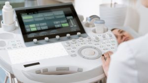
- The device performance and the quality of 3D images are highly dependent on the operator’s skill. Obtaining a good quality image requires proper application of the water-based gel and other prerequisites that can vary based on the process. For instance, an abdominal ultrasound requires a full bladder to provide good 3D image quality of the bladder and to detect stones.
Conclusion
The lack of harmful effects of ultrasound imaging, combined with its high image quality, makes it one of the best options for medical diagnosis.
The process is simple and non-invasive, making it preferable for patients and doctors. The adverse events of X-rays, such as cancer, have led researchers to utilize alternative ultrasonic techniques in almost all conventional applications of X-rays.
Even though ultrasound imaging has been prevalent for decades, technological breakthroughs are still happening. The evolution is increasing the possible quality of the 3D images and what is possible using ultrasound control pulses.
Despite the few limitations mentioned above, medical ultrasound imaging is still one of the best and safest technologies in medical science.