


There are many reasons you might have an ultrasound examination; for example, to look at a baby in your womb. It can be a little scary if you’ve never had a fetal ultrasound before or been involved in performing one. Not knowing how to interpret ultrasound images can be unsettling. However, knowing how to read an ultrasound image will put your mind at rest and make you more confident in the professional medical advice you receive.
Doctors and radiologists have lots of training on how to read ultrasound images. However, ultrasound reading isn’t too challenging once you’ve made yourself aware of the basics.
Thanks to medical imaging, the lives of millions of people have been transformed, and potentially fatal illnesses prevented. Not only are ultrasound images used during pregnancy, but also to diagnose and prevent a range of illnesses and for other therapeutic purposes.
If you’d like to know more about ultrasound images, how to read an ultrasound picture, to further your healthcare career, or because you’re a patient, keep reading.
Ultrasound Imaging Explained
Ultrasound imaging is a medical procedure that uses sound waves. The sound waves travel through the body and provide an image of the body’s internal organs and solid tissues. Such ultrasound images allow medical professionals to check the condition of different tissues, internal organs, and bodily fluids.
Doctors and other healthcare professionals use ultrasound readings to diagnose problems with a person and determine the source of pain or inflammation in the body.
Ultrasound imaging is also one of the most common testing methods when gynecologists and other healthcare professionals want to check or monitor the baby’s development.
Pregnancy ultrasound images are a popular method of testing and diagnosing because it is non-invasive and provides real-time images. An ultrasound scan is also a very safe examination method.
An ultrasound machine works using high-frequency sound waves. The waves are emitted toward the body, and they penetrate the skin. The soundwaves then bounce off a person’s tissue and inner organs.
The ultrasound machine records the reflected sound waves and the resulting patterns generate a visual image of a person’s internal organs and tissues.
The typical frequency of ultrasound waves is between 2 MHz and 15 MHz. Frequencies that are higher produce a shorter wavelength and increased attenuation. When the frequency is reduced, the absorption allows the user to study body structures and other features.
What do the Numbers Mean at the Top of an Ultrasound Image?

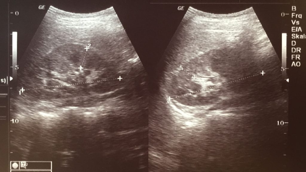
The top of an ultrasound image usually shows a series of numbers and other information. Ultrasound centers and hospitals tend to use this space for details such as:
- Patient name
- Hospital reference number
- Ultrasound machine settings
These numbers and details also help you recognize the top of ultrasound images. However, it’s not always the case. The top of an ultrasound image depends on where the ultrasound probe is placed. You’ll be able to recognize a cone-like shape in the picture. The ultrasound picture fans out from a small to a large section. The smaller end is the top of the ultrasound image.
What do different colors on an ultrasound mean?
Different colors are showcased depending on how the sound waves reflect off a substance. Most images are black and white, but you’ll notice different shades in an ultrasound scan. The different shades represent the sound waves passing through or reflecting off material densities.
Traditionally, ultrasound images were black, white, and grayish shades. A breakthrough in medical ultrasound was the advent of the color Doppler. Using this new technique, healthcare professionals were able to directly observe the flow of blood, particularly within the heart. The colors represent the direction and speed of blood flow within a specific area.
What does black on an ultrasound mean?
The image will be darker if tissues are filled with liquid, such as amniotic fluid in the uterus.
What does white mean on an ultrasound?
White on an ultrasound indicates the presence of bone.
Grayish Colors – Tissues

Grayish colors indicate tissue and liquids. The brighter the shade of gray, the denser the tissue.
What does the color orange mean on an ultrasound?
Orange is possible when using power Doppler ultrasound machines. It is used when analyzing slow flow. If you see the color orange, it generally indicates blood vessels.
What does red or blue mean on an ultrasound?
Red and blue colors represent the movement of the blood. Blue represents blood flow away from the probe, while red represents the blood flowing towards the probe.
If the screen shows a combination of blue and red, it could indicate circular flow, coherent flow, or turbulence.
Different shades of blue and red indicate velocity. Lighter shades show higher velocity, and darker shades indicate a lower velocity.
What is Shown at the Top of the Ultrasound Image?
If you ignore the numbers and text that appear at the top of the screen or printed ultrasound picture, the top of the screen or printed image is where the ultrasound probe was placed.
For example, if the ultrasound is of a uterus, at the top, you’ll see an outline of the tissues present in that area. Moving further down the screen, it shows the deeper tissue along with the uterus lining and amniotic fluid. With a fetal ultrasound, the top looks like lots of dense tissue, which corresponds with the top of the uterus and the tissue above it. Immediately under this, there is generally a black area, which is the amniotic fluid.
What Are Some Common Visual Effects?
An ultrasound picture doesn’t provide the clearest of images. It’s often the case that sound waves don’t bounce off surfaces in the body evenly. Because of this, visual effects can be used that rely on the settings, density, and angles set by the doctor or radiologist. Some of the most common visual effects are:
- Enhancement
- Attenuation
- Anisotropy
Enhancement
An enhancement is when sections of the structure that are being examined look brighter than expected. This could be due to excess fluid in the area; for example, if there is a cyst.
Attenuation
This effect is also known as shadowing and makes the area scanned appear darker than expected.
Anisotropy
This visual effect is related to the probe angle. For example, when the probe is held at right angles, it can make the area look brighter. This is particularly relevant when sound waves hit tendons. The probe angle must be readjusted to prevent it so that it contacts the body perpendicularly.
How to Understand Which Side of Your Body is Left and Which is Right?
Not all ultrasound equipment produces the same kind of image. Some produce a mirror image and others do not. However, it’s relatively easy to determine whether you’re looking at a mirrored ultrasound image or one that’s a straight shot.
Understanding the difference means you’re not going to be confused about which side of the image is the left side of your body and which is the right. Of course, you can always ask the ultrasound technician for clarification.
Generally, most ultrasound images produced in clinical settings have a mirror-like effect. In other words, what you see on the left of the ultrasound screen or ultrasound picture is the left side of the body.
However, there are clinical ultrasounds that use straight-shot imaging instead. One of the most common is transvaginal ultrasound imaging. In this situation, the left side of the body is on the right side of the ultrasound screen.
Knowing How to Read Pregnancy Ultrasounds

Ultrasound procedures are performed for a variety of reasons. One of the most common applications is checking the development of a baby during pregnancy. Ultrasound tests are very safe during such an important stage in a person’s life because, unlike X-rays, they don’t penetrate bones. Rather than use radiation, ultrasounds use sound waves or echoes to create images of the fetus or baby.
An ultrasound image reading can be very helpful at various stages of pregnancy:
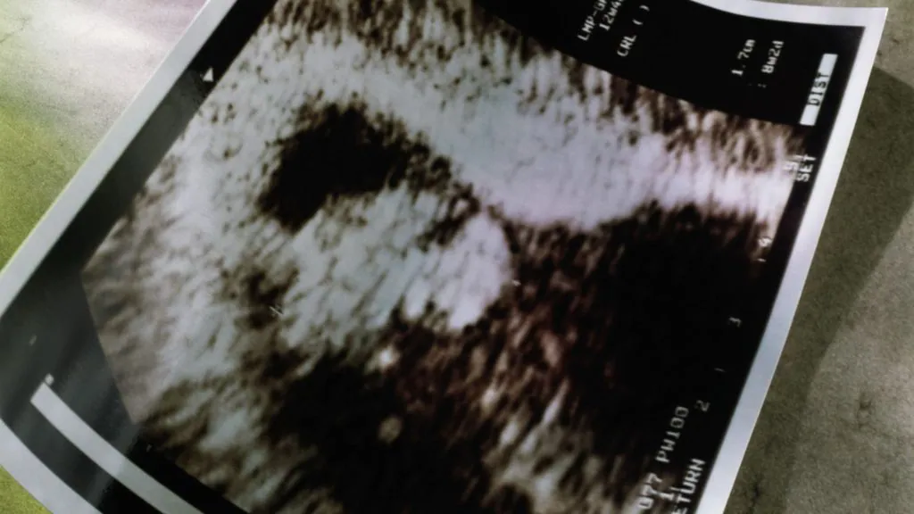
- First trimester: Within the first three months of pregnancy, an ultrasound is used to check the growth of an embryo, confirm the number of embryos, check the amniotic fluid, and calculate gestational age and estimated date of delivery (EDD).
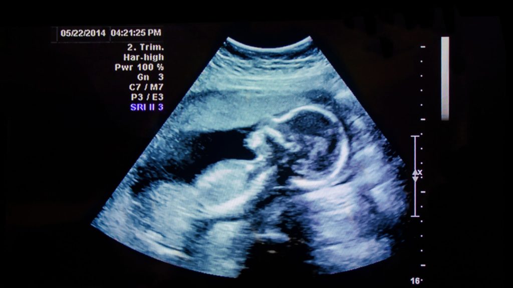
- Second trimester: Ultrasounds taken during weeks 18 and 20 allow an ultrasound technician to check the development of the fetus and take fetal measurements, its limbs, spine, internal organs, baby’s head, and brain. It’s also possible to check the location and size of the placenta, the gestational age of the fetus, and a baby’s gender if the parents want to know.
- Third trimester: Ultrasounds after week 30 of a pregnancy can be used to determine the baby’s average continued growth. Checks can be made on the location of the placenta to ensure there are no problems with it blocking the cervix.
Locate Your Womb
If you’re performing or looking at an ultrasound during pregnancy, one of the first things you should know is where the womb is.
It’s easy to locate the womb by looking at the light gray or white line around the outside of the ultrasound image. Within those lines, there will be a large black area. This indicates the amniotic fluid.
Depending on how the ultrasound doctor has positioned the transducer, your womb may or may not take up the whole sonogram image.
Locate Your Baby
The next thing to notice during ultrasound interpretations is the baby. It will look either gray or white on the image. It will be located inside the darker area of the image, which is the amniotic fluid. The amount of detail you can see will depend on the pregnancy state and the development of the baby. Some of the things you might be able to recognize include:
- 8th week of pregnancy: You’ll see an image of a fetus that looks around the same size as a baked bean.
- 12th week of pregnancy: The ultrasound image will clearly show the baby’s head.
- 20th week of pregnancy and later: You’ll be able to recognize the baby’s feet, heart, eyes, spine, and much more.
How can you tell gender on an ultrasound picture?
If you want to know your baby’s sex, you’ll be able to find this out with an ultrasound between 18 and 20 weeks of pregnancy. Ultrasound technicians look for three lines that signify a penis or labia.
Some ultrasound signs are gender-specific.
Girl ultrasound signs
- Hamburger sign: A girl’s labia lips look similar to a hamburger bun, while the clitoris resembles a hamburger patty.
- Sagittal sign: Each sex has a different sagittal sign. You can find this sign by looking at a profile view of the fetus, otherwise known as the midline sagittal plane. At the end of the spine, you’ll see a nub, the caudal notch. It indicates a girl if it points downward at a 10-degree angle.
Boy ultrasound signs
- Sagittal sign: If the caudal notch points upwards at more than a 30-degree angle, it indicates the fetus is male.
- Urine flow: Sometimes, it’s possible to spot urine flow. If the flow is upward, it is more likely the fetus is a boy.
- Male genitalia: By weeks 18 and 20, male genitalia can sometimes be seen. For example, you might be able to see the penis, scrotum, and testicles.
Traditional ultrasound is a good technique for determining a baby’s sex, but not 100% accurate. For example, some visual effects could interfere with ultrasound interpretations.
Conclusion
Now you know a little bit more about how to read an ultrasound, you won’t feel so nervous when undertaking such an examination, whatever side of the machine you sit on.
FAQs
Does cancer show up white or black on an ultrasound picture?
Cancerous tissue appears black during an ultrasound, whereas dense tissue is much lighter.
What does the CM mean on an ultrasound picture?
In a pregnancy scan, ‘cm’ usually refers to the crown-rump length, in centimeters (cm)
What does shadow on ultrasound mean?
A shadow simply means that the sound waves were reflected. It can be caused by many things, such as calcifications, air, and very dense masses or tissue.