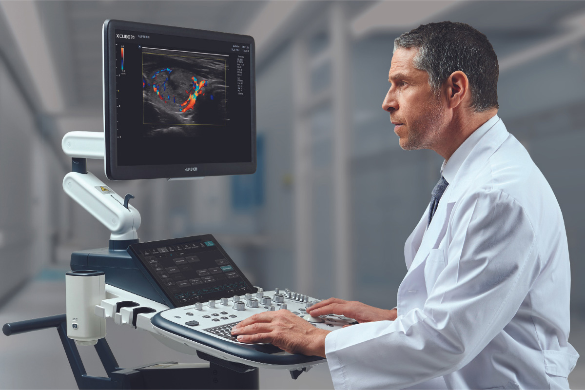
The Alpinion X-Cube 70 focuses on the needs of healthcare providers to help the provide a higher quality of patient care. The X-Cube 70 features Alpinion’s premium imaging platform, X+ Architecture, with high resolution, high sensitivity, wideband transducers and software for various applications.
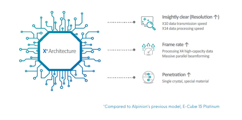
Powered by X+ ArchitectureThe Premium Imaging Platform
X+ Architecture is a premium-grade imaging platform created by combining X+ Crystal signature™, the transducer technology with high sensitivity, wideband, and X+ FIT, ALPINION’s cutting edge beamforming and data processing technology. X+ FIT is a new technology that transmits ultrasound beam width sharply, receives a large volume of data and processes it at a high speed.
In addition, X+ Crystal signature™ applies a single crystal and Alpinion’s own special material in the transducer to achieve excellent clarity to extend insight.
Expanded Capability
The Alpinion X-CUBE 70 is designed to provide an objective and accurate diagnoses based on expanded technology.
Intelligent clinical features upgrade diagnostic accuracy to new levels. Broaden your skills and range of capabilities.
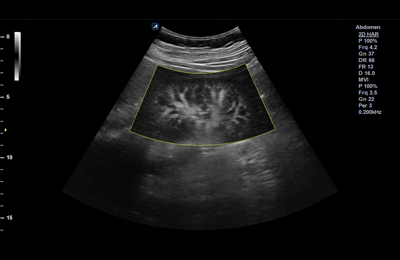
X+ MicroView
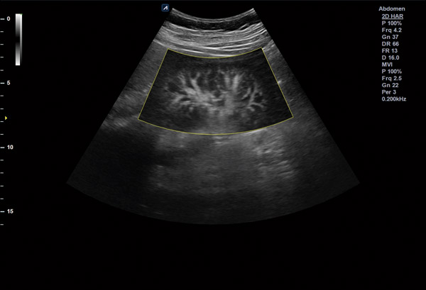
MicroView is the vascular imaging mode which displays micro blood flow. Users can observe the low-speed blood flow of tiny blood vessel. With this technology, low-speed blood flow areas which users previously couldn’t see on Color Doppler are shown at the fast frame rate.
X+ pSWE
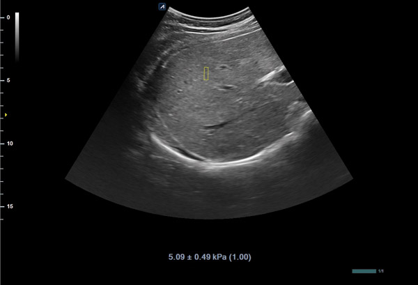
Point Shear Wave Elastography shows objective quantified stiffness of tissue using the radiation force from a focused ultrasound beam. Users can identify significant liver fibrosis non-invasively, so it reduces unnecessary biopsies and increases patient satisfaction. X+ pSWE ensures the highest reliability of measured results by displaying the Reliability Index.
CEUS (Contrast Enhancement Ultrasound)
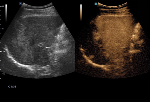
This is a function to diagnose patients using various angiographic patterns that appear while a contrast medium, administered intravenously, diffuses in blood vessels and organ tissue. CEUS has many advantages in various clinical indications for liver disease.
GENERAL IMAGING
X+ MicroView
Microview is the vascular imaging mode which displays micro blood flow. Users can observe the low-speed blood flow of tiny blood vessels. With this technology, low-speed blood flow areas which users previously could not see on Color Doppler are shown at a fast frame rate.
X+ pSWE
Point Shear Wave Elastography shows objective quantified stiffness of tissue using the radiation force from a focused ultrasound beam. Users can identify significant liver fibrosis non-invasively, so it reduces unnecessary biopsies and increase patient satisfaction. X+ pSWE especially ensures the highest reliability of measured results by displaying the Reliability Index.
CEUS (Contrast Enhancement Ultrasound)
CEUS is a function to diagnose patients using various angiographic patterns that appear while a contrast medium, administered intravenously, diffuses in blood vessels and organ tissue. CEUS has many advantages in various clinical indications for liver disease.
DPDI (Directional Power Doppler Imaging)
The technology brought improvement in delivering information related to direction of blood flow and sensitivity of Color Doppler. This is useful in sensing relatively slower blood flow in peripheral blood vessels (e.g., renal vessels, peripheral vessels and middle cerebral artery).
MSK
Needle VisionTM Plus
Beam steering displays the shape of a needle and in which direction the needle is heading to safely guide injection. With this needle enhancement function, accuracy and safety of procedures will be improved using a linear transducer. Beam angles can be adjusted by three levels so that direction and shape of the needle are visible with advanced clarity.
Elastography
Elastography is ultrasound imaging technology that reveals relative elasticity of tissues against external pressure. In parallel, relevant pathological information is offered to help reduce unnecessary biopsy. Pressure level on tissues can be monitored in real time with an indication bar marked with its scale of one (1) to six (6) to enhance the credibility of results.
Panoramic
Panoramic Imaging offers a horizontal image with an extremely extended field of view.
WOMEN'S HEALTH
Silhouette Imaging
This new technology provides silhouette outlines of the human fetus and other structures clearly. By using Live HQTM technology with a shadowing effect. Silhouette imaging technology allows to delineate the outlines of structures present behind the directly visualized structure
Live HQ
The combination of qualified 3D/4D transducers with strong performance reliability and geometrical accuracy enables Live HQTM software to render a realistic fetal shape.
Elastography
Elastography is ultrasound imaging technology that reveals relative elasticity of tissues against external pressure. In parallel, relevant pathological information is offered to help reduce unnecessary biopsy. Pressure level on tissues can be monitored in real time with an indication bar marked with its scale of one (1) to six (6) to enhance the credibility of results.
Depth View
Depth View is a rendering mode that gives a three-dimensional effect by applying color according to the depth in direction depth form the view point.
Clear Face
Clear Face detects a fetal face automatically and it removes structures that cover the fetal face such as the cord, placenta and uterus. It allows users to acquire clear view with simple operation.
CARDIOVASCULAR
Auto EF
Auto EF evaluates and measures contraction of the left ventricle by automatically calculating Ejection Fraction (EF). In addition to EF, this function also provides ESD and ESV information to display volume change of the left ventricle. Data can be contained without user control by automatically detecting the boundary line of the endocardium.
CUBE StrainTM
With Alpinion’s phased array transducers implemented with X+ Crystal Signature technology, more clarity in images is achieved, and with use of the functions, early diagnosis of cardiac disorders can be made possible.
Stress Echo
Stress Echo offers an accurate diagnosis of cardiac disorders by quantifying myocardial deformation.
Auto IMT
Auto IMT allows you to automatically measure intima-media thickness of carotid arteries. It enables more reliable and reproducible assessment.
Clinical Images
Alpinion X-Cube 70 Workflow
X+ Assistant
X+ Assistant enables users to reduce key strokes by at least 50% and save time when conducting examinations. In addition, it is also possible to register applications and protocols optimized.
X+ Auto Biometry
When measuring Estimated Fetal Weight(EFW),
the smart recognition algorithm allows you to automatically identify a structure of interest and measure fetal head circumference (HC), biparietal diameter(BPD), femur length (FL), abdominal circumference (AC) and Humerus.
X+ Compare
This is a feature that allows users to import patients’ previous
study from a PACS server or hard disk and compare when scanning and reviewing. X+ Compare can be used not only in scan mode, but in review mode (E-View) as well. * X+ Compare supports ultrasound studies only.
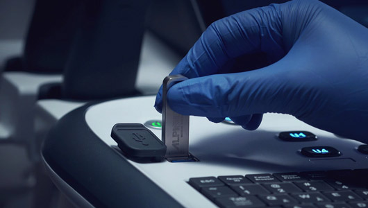
USB Real-Time Recording
USB real-time recording makes data transfer easier by allowing users to record ultrasound scan images on USB memory in real time. Videos are recorded as high-definition and stored in system quickly.
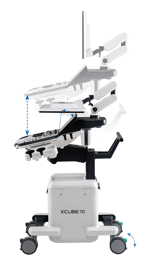
Intuitive Design
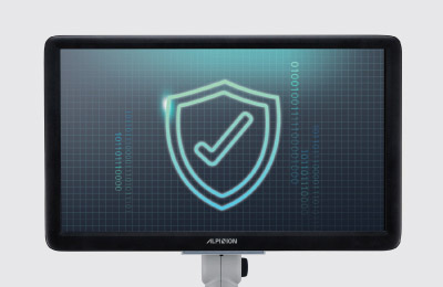
Antivirus Solution
The automatic virus scanning function is activated whenever the system boots, patients’ information is more secure and protected.
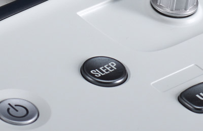
Time Saving Solution
The built-in battery guarantees mobility while it is in use. Sleep mode helps the system boot up faster.
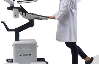
Compact Size & Weight
The X-Cube 70 is compact and light weight, measuring 85 kg providing for great mobility.
Design and Ergonomics
23 inch FHD LED Screen
12.1 inch tilting LCD Color Panel
Motorized Control Panel
5 Transducer Connectors
4 Swivel Wheels
Swivel Lock
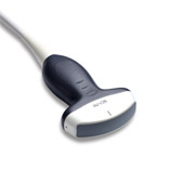
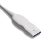
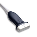
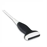
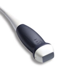
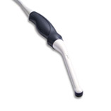
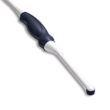
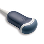
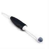
Transducer Technology
The key to transducer technology is its process of generating a high sensitivity, wideband acoustic signal and receiving it without any loss, and then converting it to a digital signal. ALPINION MEDICAL SYSTEMS has researched and developed the core technology of transducers and X+ Crystal Signature is the accomplishment of all the innovative technologies they have accumulated.
@ Copyright 2023 Ultrasound Solutions Corp. | All Rights Reserved Sitemap | privacy Policy | Equipment Quotation - Terms & Conditions | Our Sister Company Hmiixraysales
| Powered by Dipoletechi
Log In
Schedule A Demo
Ready to schedule a demo or have more questions? We are here to help.
Our sales team is here to help you choose your next ultrasound machine. They can answer all of your questions and set up your demo, just fill out the form and we will be in touch ASAP.


 X+ MicroView
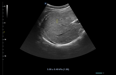 X+ pSWE
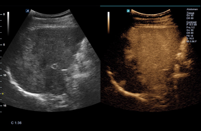 CEUS (Contrast Enhancement Ultrasound)
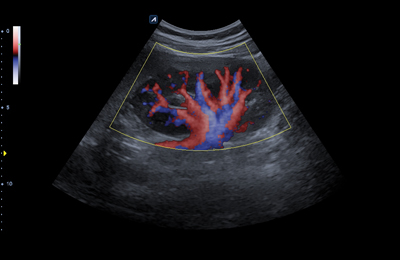 DPDI (Directional Power Doppler Imaging)
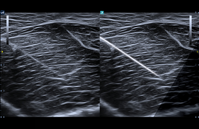 Needle VisionTM Plus
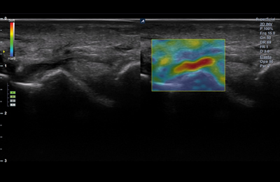 Elastography
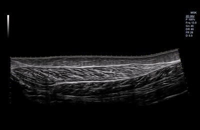 Panoramic
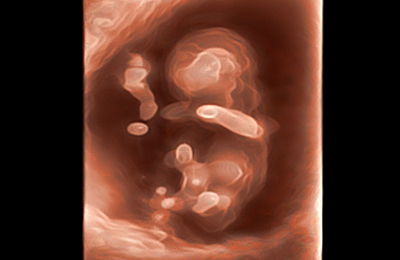 Silhouette Imaging
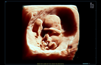 Live HQ
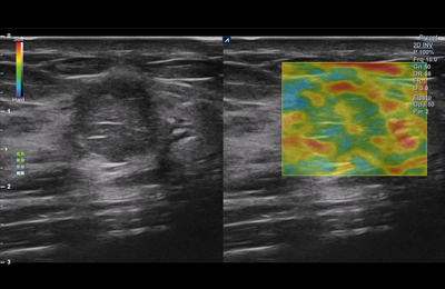 Elastography
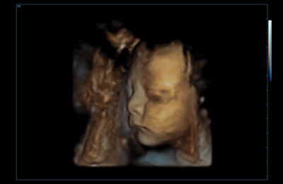 Depth View
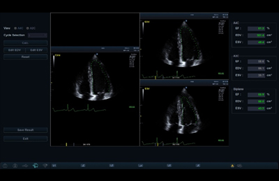 Auto EF
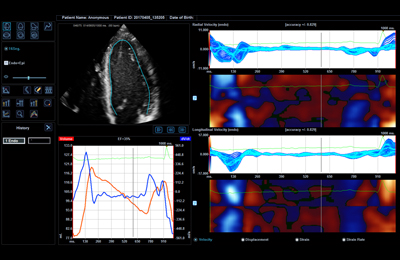 CUBE StrainTM
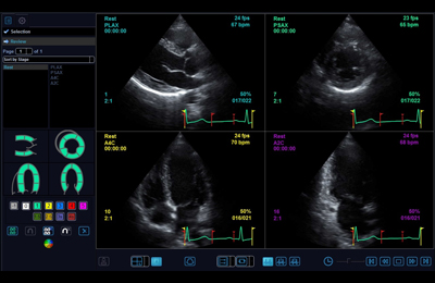 Stress Echo
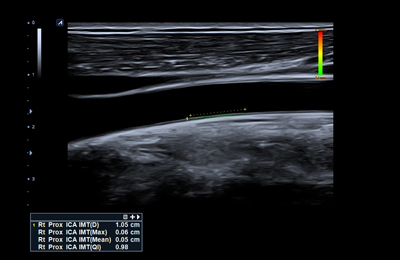 Auto IMT