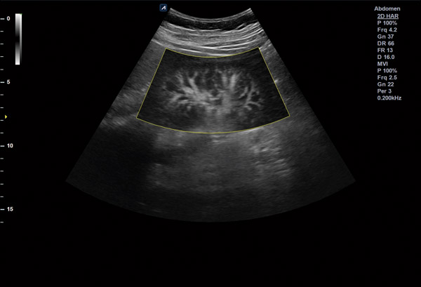
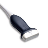
 X+ MicroView
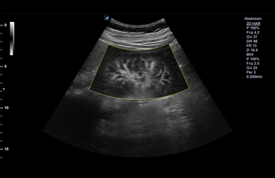
Microview is the vascular imaging mode which displays micro blood flow. Users can observe the low-speed blood flow of tiny blood vessels. With this technology, low-speed blood flow areas which users previously could not see on Color Doppler are shown at a fast frame rate.
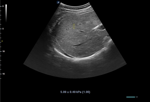
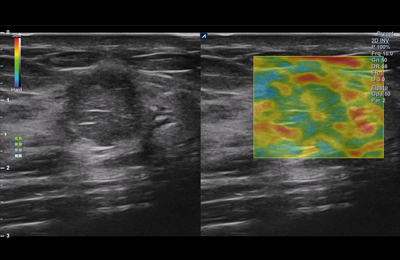
 X+ pSWE
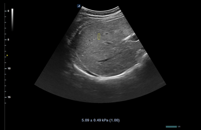
Point Shear Wave Elastography shows objective quantified stiffness of tissue using the radiation force from a focused ultrasound beam. Users can identify significant liver fibrosis non-invasively, so it reduces unnecessary biopsies and increase patient satisfaction. X+ pSWE especially ensures the highest reliability of measured results by displaying the Reliability Index.
 CEUS (Contrast Enhancement Ultrasound)
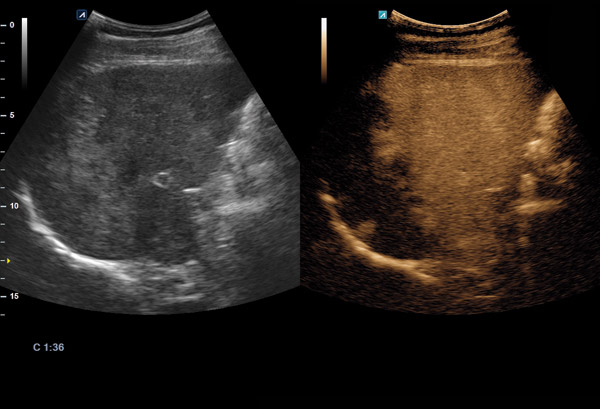
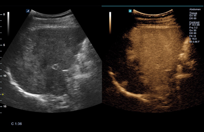
CEUS is a function to diagnose patients using various angiographic patterns that appear while a contrast medium, administered intravenously, diffuses in blood vessels and organ tissue. CEUS has many advantages in various clinical indications for liver disease.
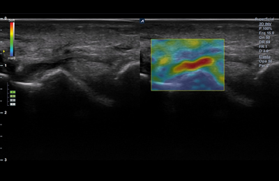
 DPDI (Directional Power Doppler Imaging)
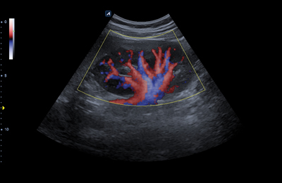
The technology brought improvement in delivering information related to direction of blood flow and sensitivity of Color Doppler. This is useful in sensing relatively slower blood flow in peripheral blood vessels (e.g., renal vessels, peripheral vessels and middle cerebral artery).
 Needle VisionTM Plus
Beam steering displays the shape of a needle and in which direction the needle is heading to safely guide injection. With this needle enhancement function, accuracy and safety of procedures will be improved using a linear transducer. Beam angles can be adjusted by three levels so that direction and shape of the needle are visible with advanced clarity.
 Elastography
Elastography is ultrasound imaging technology that reveals relative elasticity of tissues against external pressure. In parallel, relevant pathological information is offered to help reduce unnecessary biopsy. Pressure level on tissues can be monitored in real time with an indication bar marked with its scale of one (1) to six (6) to enhance the credibility of results.
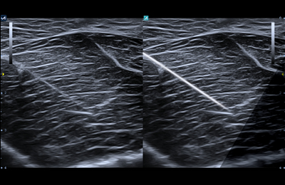
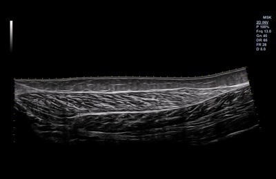 Panoramic
Panoramic Imaging offers a horizontal image with an extremely extended field of view.
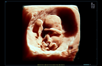
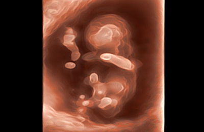 Silhouette Imaging
This new technology provides silhouette outlines of the human fetus and other structures clearly. By using Live HQTM technology with a shadowing effect. Silhouette imaging technology allows to delineate the outlines of structures present behind the directly visualized structure
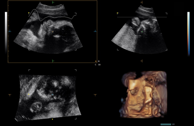
 Live HQ
The combination of qualified 3D/4D transducers with strong performance reliability and geometrical accuracy enables Live HQTM software to render a realistic fetal shape.
 Elastography
Elastography is ultrasound imaging technology that reveals relative elasticity of tissues against external pressure. In parallel, relevant pathological information is offered to help reduce unnecessary biopsy. Pressure level on tissues can be monitored in real time with an indication bar marked with its scale of one (1) to six (6) to enhance the credibility of results.
 Depth View
Depth View is a rendering mode that gives a three-dimensional effect by applying color according to the depth in direction depth form the view point.
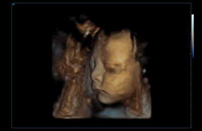
 Clear Face
Clear Face detects a fetal face automatically and it removes structures that cover the fetal face such as the cord, placenta and uterus. It allows users to acquire clear view with simple operation.
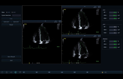 Auto EF
Auto EF evaluates and measures contraction of the left ventricle by automatically calculating Ejection Fraction (EF). In addition to EF, this function also provides ESD and ESV information to display volume change of the left ventricle. Data can be contained without user control by automatically detecting the boundary line of the endocardium.
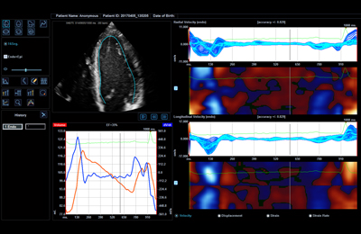 CUBE StrainTM
With Alpinion’s phased array transducers implemented with X+ Crystal Signature technology, more clarity in images is achieved, and with use of the functions, early diagnosis of cardiac disorders can be made possible.
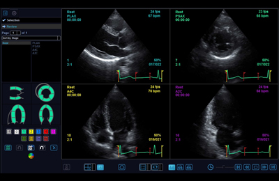 Stress Echo
Stress Echo offers an accurate diagnosis of cardiac disorders by quantifying myocardial deformation.
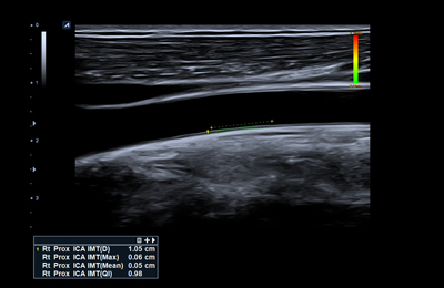 Auto IMT
Auto IMT allows you to automatically measure intima-media thickness of carotid arteries. It enables more reliable and reproducible assessment.