Diverticular disease accounts for approximately 312,000 hospital admissions in the United States annually, and costs nearly $2.6 billion dollars. Diverticulitis is almost unheard of in the developing world where people general eat “high-fiber” diets, but in America the disorder is becoming more common. Approximately 20% of Americans with diverticular disease will experience at least one episode of acute diverticulitis, necessitating a visit to their physician’s office or the emergency room for treatment.
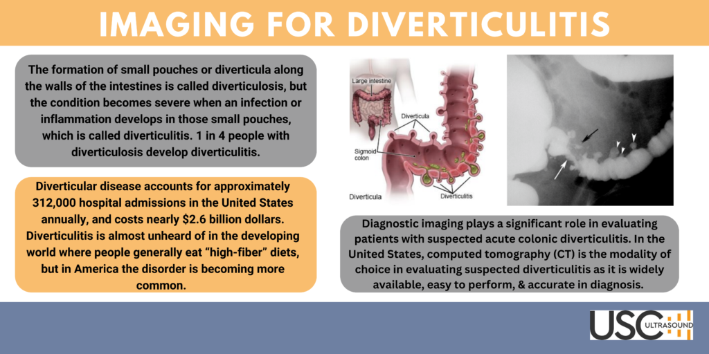
 The formation of small pouches or diverticula along the walls of the intestines is called diverticulosis, but the condition becomes severe when an infection or inflammation develops in those small pouches, which is called diverticulitis. 1 in 4 people with diverticulosis will develop diverticulitis. In some serious cases, diverticulitis may lead to a severe perforation of the bowel. The pouches are more common at the end of the sigmoid colons, which is on the left side of the abdomen, but they can form anywhere on the digestive tract.
The risk of developing diverticulitis rises with age (nearly 50% of people aged over 60 years have colonic diverticula). As the population of the United States continues to age, this larger population group is contributing to the growing number of cases of diverticulitis.
Diverticulitis is usually diagnosed during an acute attack. Because abdominal pain can indicate a number of problems, a physician will need to rule out other causes for the patient’s symptoms. One of the main symptoms of diverticulitis includes abdominal pain and cramping that is more severe on the left side. Other common symptoms include chills, nausea, fever, rectal bleeding, and constipation or diarrhea.
How is Diverticulitis Diagnosed?
Typically, a healthcare provider will start with a physical examine. He or she will ask questions about the patient’s symptoms and health history. Further assessment will usually include:
- Blood and urine tests may show signs of infection or inflammation.
- CT scan or ultrasound images may be used to find problems in within the intestines. A contrast liquid maybe used to help the intestines show up better in the images.
- A colonoscopy is used to look at the intestines with a scope. A scope (long bendable tube with a light on the end) is used to take additional images. This test may show swollen diverticula or bleeding. Samples may be taken from the digestive tract and sent to a lab for tests. Bleeding may be controlled with tools that are inserted through the scope.
Diagnostic imaging plays a significant role in evaluating patients with suspected acute colonic diverticulitis. The diagnosis can be confirmed by imaging, and a combination of the location of the disease, severity of inflammation, and complications will allow clinicians to determine appropriate treatment. In the United States, computed tomography (CT) is the modality of choice in evaluating suspected diverticulitis as it is widely available, easy to perform, and accurate in diagnosis. Although CT imaging is considered the “gold standard” for the diagnosing diverticulitis in America, ultrasound is the preferred option in Europe, Asia, and Africa as the initial imaging modality in the evaluation of patients with suspected diverticulitis. Recent studies have suggested that there is no significant difference in the test performance characteristics of CT as opposed to ultrasound for the diagnosis of diverticulitis.
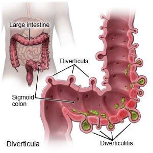
CT Image of Diverticulitis with Abscess
Findings:
-
- White arrowheads: Diverticula
- White arrow: Narrowed lumen
- Black arrow: Perforation with intramural abscess
While CT has long been the diagnostic modality of choice to confirm acute diverticulitis in the United States, given the high cost associated with CT imaging, many countries utilize ultrasound as the first-line imaging study to make this diagnosis.
When evaluating the patient with ultrasound the clinician or sonographer performs an abdominal ultrasound to assess for diverticulitis by gently sweeping through loops of the bowel in the area of localized abdominal pain. A low-frequency curvilinear array transducer is used to visualize bowel as it provides adequate depth. The clinician can ask the patient to point to the area of maximal tenderness, which often indicates the highest-yield area for sonographic evaluation. The sonographer evaluates the symptomatic region with the transducer, looking for evidence of acute diverticulitis: (1) at least one diverticulum; (2) thickening of the bowel wall (typically 4–5 mm or more); and (3) echogenic non-compressible fat surrounding one or more diverticula, suggesting an acute inflammatory process. In addition, the sonographer may identify a so-called “target sign” or “pseudokidney sign”, which refers to the hypoechoic wall surrounding a hyperechoic center
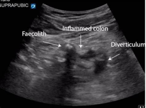
What are the imaging findings of diverticulitis?
-
- US: Abnormal wall thickening of more than 4 mm involving a segment 5 cm or longer at the point of maximal tenderness.
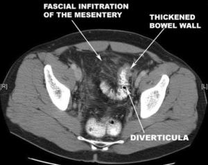
- CT:
- Diverticula
- Narrowed lumen
- Thickened bowel wall
- Fascial inflammatory infiltration
- Complications
- Perforation: Free air in the peritoneum
- Abscess
If utilized by providers with appropriate training, point-of-care ultrasound represents an ideal first-line imaging test for uncomplicated diverticulitis, with CT imaging reserved for ill-appearing patients with unstable vital signs, or for those whose point-of care ultrasound images demonstrate findings concerning complicated diverticulitis, such as an abscess or intra-abdominal free fluid.
Treatment
Uncomplicated Diverticulitis
- Antibiotics to treat infection, although new guidelines state that in very mild cases, they may not be needed.
- A liquid diet for a few days while the bowel heals. Once the patient’s symptoms improve, gradually add solid food to the patient’s diet.
Complicated diverticulitis
- Intravenous antibiotics
- Insertion of a tube to drain an abdominal abscess, if one has formed
Surgery
If:
- complication, such as a bowel abscess, fistula or obstruction, or a puncture (perforation) in the bowel wall
- multiple episodes of uncomplicated diverticulitis
- weakened immune system
Follow Up
The Clinician may recommend colonoscopy six weeks after recovery from diverticulitis. There doesn’t appear to be a direct link between diverticular disease and colon or rectal cancer. But colonoscopy — which is risky during a diverticulitis attack — can exclude colon cancer as a cause of the patient’s symptoms.
How USC is Connected to Bringing Awareness To Diverticulitis
Recently one our Field Service Engineers was diagnosed with Diverticulitis. This occurred during an install of a Claris CT machine at an Ultrasound customers facility. I was in contact with our Field Service Engineer, Jason Scamara while he was performing the equipment installation in Las Vegas. This was an important customer and an important installation. I was monitoring the progress and both texting and calling Jason throughout the day to see how the work was coming along. Late in the afternoon Jason disclosed to me that he was in severe pain, with nausea and fever. We agreed that he would go to the doctor and get his condition looked at.
 It turned out to be diagnosed as an acute attack of Diverticulitis. Jason was given antibiotics and an IV. He spent the night at a hotel and completed the installation work the following day before making the several hour drive to his home in Lake Peris California.
I have personally known Jason since 2007. At that time, I was with Numark Industries and based at our Engineering Design office in Culver City, California. I was the Global Director of Quality and had my staff spread out in facilities across the globe. Jason was one of my team working at our Buena Park California Service and Warranty Center.
One of a handful of original DJ brands, Numark was founded in Edison, New Jersey in 1971. At the time, Numark developed a significant following for its mixers, turntables, and speaker enclosures and was considered to be one of the leading manufacturers in the DJ industry during the golden age of Disco. While Jason worked for Numark by day, at night he was performing as a DJ.
In the early 2000’s, around the time I met Jason, digital media was not only changing the DJ industry, but the consumer world as well. When Apple’s iPod hit the scene, it created a tipping point in the world of digital file-based music storage and playback, establishing hard drive (and later flash) music playback as a mainstay across all industries. The iPod quickly emerged as a serious music playback platform for consumers and DJs alike, and Numark wasted no time in creating a hard drive-based tool specifically designed for DJs that validated this rapidly evolving format. DJs no longer had to bring a binder full of CDs or even a computer with them.
 In 2007, Numark and Serato released NS7, a controller that blends different eras of DJing so completely it makes them virtually indistinguishable from each other. Since then, the powerful experience offered by NS7 has defined the top tier of DJ performance, setting the standard by which all other controllers are judged. Jason played a critical role in bringing this product to market and performed all the initial testing on the first 200 units ever produced.
I left Numark to pursue other business opportunities but our paths were to cross once again, when he joined USC as a Field Service Engineer. As USC was looking to expand into the west coast market and establish a facility in Las Vegas, Jason reached out to me and there was an opportunity available to have him join our team as a regional Field Service Engineer. Having known Jason for over 10 years, and I can say that he is a good man, a great electronic technician, and a good friend. Currently, it is still be determined between Jason and his doctor if he will need surgery for his condition. The next steps in his treatment include having a colonoscopy performed for further examination and to rule out colon cancer.
On behalf of myself and all of the USC family we wish Jason a speedy and healthy recovery.
Sources:
NIH, NCBI Ultrasound diagnosis of diverticulitis
Michael E. Abboud, Sarah E. Frasure, and Michael B. Stone
Mayo Clinic
Diverticulitis care at Mayo Clinic